
Introduction
Ankle fractures are one of the most common types of injuries seen by orthopedic surgeons, and the lateral malleolus is involved in the majority of these fractures.1 Pilon fracture complexes and distal tibia intraarticular fractures are unstable fractures that frequently involve the distal fibula and can require surgical fixation. The traditional method of repair has been open reduction and internal fixation (ORIF) with plates and screws; however, this carries a risk of wound complications like infection and soft tissue irritation in 26-40% of patients.2 In addition, hardware failure and symptomatic implants contribute to a need for additional surgery in 30% of patients.3 While stability is beneficial, these complications have led to some controversy regarding whether plate fixation is the ideal procedure. Another method utilized for patients with distal fibula fractures is intramedullary fixation, which requires less invasive surgical exposure. This is not a new technique, and various implants have been used, including wires and locked intramedullary nails.4 Intramedullary screws are also an attractive option, and here we describe intramedullary fixation of the fibula with a long cortical screw which we believe is a relatively straightforward, inexpensive, and available technique.
Indications and Contraindications
General indications for intramedullary screw fixation of distal fibula fractures are length-stable fractures in associated tibial pilon fracture patterns and very distal extraarticular tibia and fibula fractures in patients with or without high risk for wound complications and infection. Extended indications include isolated lateral malleolar, bimalleolar, and trimalleolar fractures in high-risk patients. High-risk patients include those with high-energy fractures, diabetes, peripheral neuropathy, body mass index (BMI) > 30, smokers, and the elderly. These patients are at increased risk for complications from plate fixation because the soft tissue envelope is more fragile.5 In high-risk patients, intramedullary fixation may be preferred over plate fixation. A study of 37 patients with these preexisting comorbidities found that retrograde intramedullary screw fixation was a viable alternative to plate fixation with low complication rates.5
General contraindications include malleolar fractures requiring length and rotational control in low-risk patients. Comminuted and length unstable fibula fractures may be contraindicated since it may be more challenging to maintain length and rotation with an intramedullary screw in these injury patterns, especially if early weight-bearing is anticipated. Pre-existing deformity of the fibula or a highly narrow medullary canal could prevent proper insertion and placement of the screw. If a percutaneous repair method has to be utilized, operation within 7 to 10 days is recommended to avoid the difficulties of achieving satisfactory reduction after the callus has formed.2
Surgical Technique
Surgical Preparation
Intramedullary fixation of the fibula is typically done in addition to fixation of other fractures in the lower leg, such as ORIF or intramedullary nailing of a distal tibia fracture. Therefore, setup and preparation for this technique are frequently done in the context of how the tibia is being fixed.
The patient is generally brought to the operating room and laid supine and flat. Prophylactic intravenous antibiotics are given, and the patient is given general anesthesia. The non-operative limb is carefully padded, and sequential compression devices are used on the contralateral extremity. The operative limb is often elevated on a foam positioner or blankets and carefully scrubbed and prepped.
Surgical Equipment
The necessary equipment for this technique includes a long 2.5 mm drill bit to establish the starting point and a 3.5 mm by 150 mm stainless steel or titanium cortical screw to fix the fibula. The screw tends to be flexible and blunted, so in some cases, it can be placed into the intramedullary canal without the drill bit establishing the entire length. Since the procedure is frequently percutaneous, fluoroscopic control is required.
Surgical Steps
Surgeons have different approaches to determining whether to fix the tibia or fibula first based on the nature of the injury. Fixing the fibula first may be more optimal if the tibia is comminuted and shortened because initial fixation of the fibula can establish the length confidently. However, fixing the tibia first would be risky because it can be challenging to estimate the length. On the other hand, fixing the tibia first may be better in different cases since it may be harder to manipulate the tibia, which is more critical, once the fibula is fixed.
In the fibular reduction stage, in some cases, a closed reduction is performed if the fracture can be reduced with manipulation alone. Sometimes, an open incision is needed at the level of the fracture, and instruments are used to reduce the fracture.
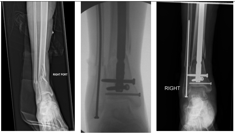
In the fibular fixation stage, the skin around the distal fibula is infiltrated with 1% lidocaine with epinephrine. A 1 cm incision over the distal fibula is made, and sharp dissection is made down to the tip of the fibula. A radiograph is used to confirm the starting point of the 2.5mm drill bit, which is then inserted in the anterolateral quadrant of the distal fibula and then directed proximally into the intramedullary canal and potentially across the fracture site. Drilling across the fracture site is not required in most instances. The drill will often tend to penetrate the medial cortex and make intramedullary screw placement difficult. In these cases, stopping short of the medial cortex is necessary. The drill is then removed, and the screw is then inserted and tends to be flexible enough to stay in an intramedullary path and then cross the fracture site without difficulty. The screw is fully seated, and care is taken not to countersink the screw head. Images are taken to confirm screw placement and satisfactory reduction of the fracture. Hemostasis is achieved, and the incision is irrigated and closed using a nylon suture. [Figures 1 and 2]


Post-Operative Management
Immediate management involves external immobilization with a splint or boot for two weeks without weight-bearing. At the end of 2 weeks, the sutures are taken out, and progressive mobilization and range of motion are recommended, given that wounds appear to be healing and no other complications have arisen. Given this technique’s potential lack of rotational stability, it is essential to assess proper healing, and additional time without weight-bearing may be necessary. Weightbearing recommendations are dependent on the nature of the corresponding tibia fracture. In the case of an extraarticular fracture, we may allow early weight-bearing. But for intraarticular distal tibia fractures or concomitant syndesmotic injuries, weight-bearing restrictions are often in place for longer.
Discussion
Plate fixation of distal fibula fractures remains the most versatile and common method and is frequently required in malleolar fracture patterns requiring anatomic reduction. However, intramedullary screw fixation of distal fibula fractures is relatively straightforward. In many cases, it can be an effective strategy for achieving stable fixation with minimal compromise of the soft tissue envelope. Patients with extraarticular very distal tibia and fibula fractures treated with tibia intramedullary nailing sometimes can fit only two distal interlocking screws in a very short segment. In these cases, simultaneous fixation of the fibula can provide needed angular stability of the construct in most fibula fracture patterns and length stability in simple fibula fracture patterns. Fixation of the fibula in tibia pilon fracture patterns is somewhat controversial due to the concern for the potential need for additional incisions or larger exposures needed in patients with compromised soft tissue envelopes. Intramedullary fibula screw fixation provides stability with minimal soft tissue compromise in these cases. Whereas multiple methods of intramedullary fixation of distal fibula fractures exist (locked intramedullary nailing, Rush pins, flexible titanium rods, Steinmann pins, etc.), fixation with a long 3.5mm cortical screw provides several advantages. Fixation with Rush or Steinmann pins can often be challenging since the cutting end of the pin tends to become impaled in the medial cortex of the fibula, as opposed to the long 3.5mm screw whose blunt tip and flexibility allow it to “find its way” and remain intramedullary. Locked intramedullary fibula nailing can provide good stability with minimally invasive methods but is far more costly than a single 3.5 mm cortical screw. [Table 1]
Intramedullary fixation was associated with significantly lower rates of wound complications, faster fracture healing, and better recovery of ankle function compared to plate fixation.6 This can be explained by the minimally invasive nature of this procedure, which decreases the risk of infection and preserves the local hematoma, which contains mediators that promote the healing process. Loukachov et al. found a wound infection rate of 0.6% and an anatomic reduction rate of 93%.3 Albana et al. also saw similar rates in patients with multiple risk factors for wound healing complications, which represent the largest demographic of patients who sustain ankle fractures.5
Studies investigating intramedullary nailing, which follows a similar concept as intramedullary screw fixation, have shown faster rates of recovery and potentially open the possibility of earlier weightbearing.7 This could be related to the biomechanical properties of the two surgical constructs. Smith et al. found that fibular nailing could withstand greater rotational torque forces in Weber B fractures and held the fibular construct better than the traditional plate.8
Post-operative complications related to intramedullary fixation were found to occur in 10.3% of patients and were mainly associated with the implant rather than wound complication. Commonly identified issues include implant failure and fibular shortening, which were related to the use of earlier designs and the use of the technique in unsuitable fracture patterns.9 This may reflect the challenges that arise from a learning curve of a new technique, and further investigation is required to optimize the hardware and understand when this procedure is most appropriate. Further implant-related complications using the 3.5mm screw include secondary displacement and nonunion, which could potentially be due to insufficient stabilization of the fibula fracture.3 In addition, the percutaneous nature of this procedure opens up the risk of injury to the peroneal tendons and sural nerve, which are close to the surgical site. Guidewire placement in the anterolateral quadrant of the distal fibula can most mitigate these concerns.10
In conclusion, intramedullary screw fixation of the fibula is a straightforward, low-cost, safe, and effective method for helping stabilize the lower leg and ankle, which should be considered by surgeons who care for these injuries.
Declaration of Conflict of Interest
SR has consulting agreements with Depuy Synthes and Globus Medical and has received royalties from Globus Medical. JK declares no competing interests related to this work.
Declaration of Funding
The authors received NO financial support for the preparation, research, authorship, and publication of this manuscript.
Declaration of Ethical Approval for Study
This study is exempt from IRB approval as patient records were not reviewed for this technique paper.
Declaration of Informed Consent
There is no information in the submitted manuscript that can be used to identify patients.