
INTRODUCTION
Hip fractures are a common orthopaedic injury in the elderly population and can lead to substantially reduced quality of life.1,2 Displaced femoral neck fractures are frequently treated with either cemented or uncemented hemiarthroplasty, with each conveying its own unique benefits and drawbacks.3 Cemented hemiarthroplasty has been shown to result in better quality of life than uncemented hemiarthroplasty.4 This technique is also associated with lower incidence of periprosthetic fracture, prosthetic subsidence, and implant loosening.4,5 Conversely, cemented hemiarthroplasty is correlated with reduction in intraoperative blood pressure and, although rare, can result in cardiovascular collapse and death.4 Furthermore, it has been reported that approximately half of the mortalities that occur within one day of cemented hemiarthroplasty can be attributed to the use of bone cement, known more formally as bone cement implantation syndrome.6
In contrast, uncemented hemiarthroplasty allows for a shorter operation time and less procedural blood loss in comparison to cemented hemiarthroplasty.6 Additionally, one investigation reported a reduced risk of postoperative pulmonary embolism following uncemented hemiarthroplasty.7 However, it has been reported on numerous occasions that uncemented hemiarthroplasty conveys a higher risk of periprosthetic fracture.4,5,8,9 Fernandez et al. reported a more than four-fold increased prevalence of periprosthetic fractures in uncemented hemiarthroplasty (2.1%) compared with cemented hemiarthroplasty (0.5%).4 This complication is particularly devastating, as periprosthetic fracture following hemiarthroplasty was associated with a 30 day mortality rate of 12.5% and a one year mortality rate of 28.1%.10
Based on these data, it is important to consider the risk of periprosthetic fracture if opting for uncemented hemiarthroplasty. One potential factor that may influence the rate of periprosthetic fracture following hemiarthroplasty is proximal femur morphology, which can be described via the Dorr classification.11 This system classifies patients into one of three categories: Dorr A, B or C. Dorr A patients possess thick and distinct cortices with a narrow diaphyseal canal.12 This is followed by Dorr B patients who have a thinner cortex and wider diaphyseal canal, followed by Dorr C patients who demonstrate substantial loss of medial and posterior cortices alongside a very wide canal.12 Ultimately, this morphology can affect femoral stem fit and bone quality, particularly following hemiarthroplasty, which may translate to worsened postoperative outcomes.12 Alternatively, cortical thickness index (CTI) is a measure of underlying bone mineral density (BMD) that may play a role in perioperative outcomes following hemiarthroplasty, notably intraoperative periprosthetic fracture.13,14 However, it remains unclear whether decreased CTI is associated with periprosthetic fracture after successful implant placement.15,16 The purpose of this study was to further investigate the association between both Dorr classification proximal femur morphology, cortical thickness index, and the incidence of periprosthetic fracture following uncemented hemiarthroplasty. Our hypothesis is that patients with Dorr C morphology will have a higher prevalence of periprosthetic fracture than those with Dorr A and Dorr B morphology whereas CTI will demonstrate a negative correlation with this outcome.
METHODS
Institutional Review Board approval was obtained, and a retrospective chart review was conducted of patients who underwent hemiarthroplasty for femoral neck and head fractures at a single level one trauma center from January 1, 2011 to December 31, 2021. Only patients who underwent uncemented hemiarthroplasty were included. In total, there were 97 patients diagnosed with a femoral neck and head fracture who underwent an uncemented hemiarthroplasty (CPT 27236) during this period. Adult patients (ages 18 and older) were included in the study. Prisoners, pregnant individuals, and patients with concurrent injuries were excluded.
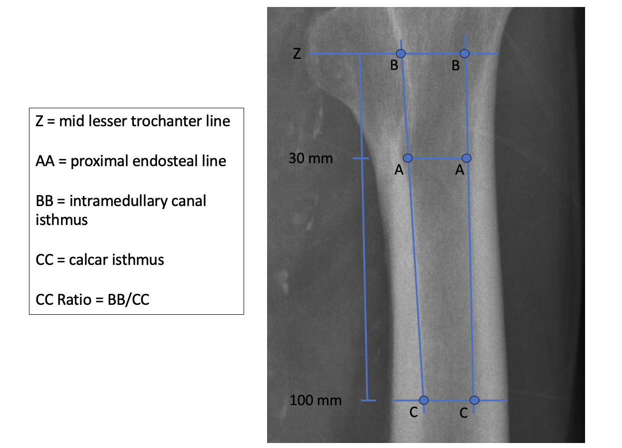
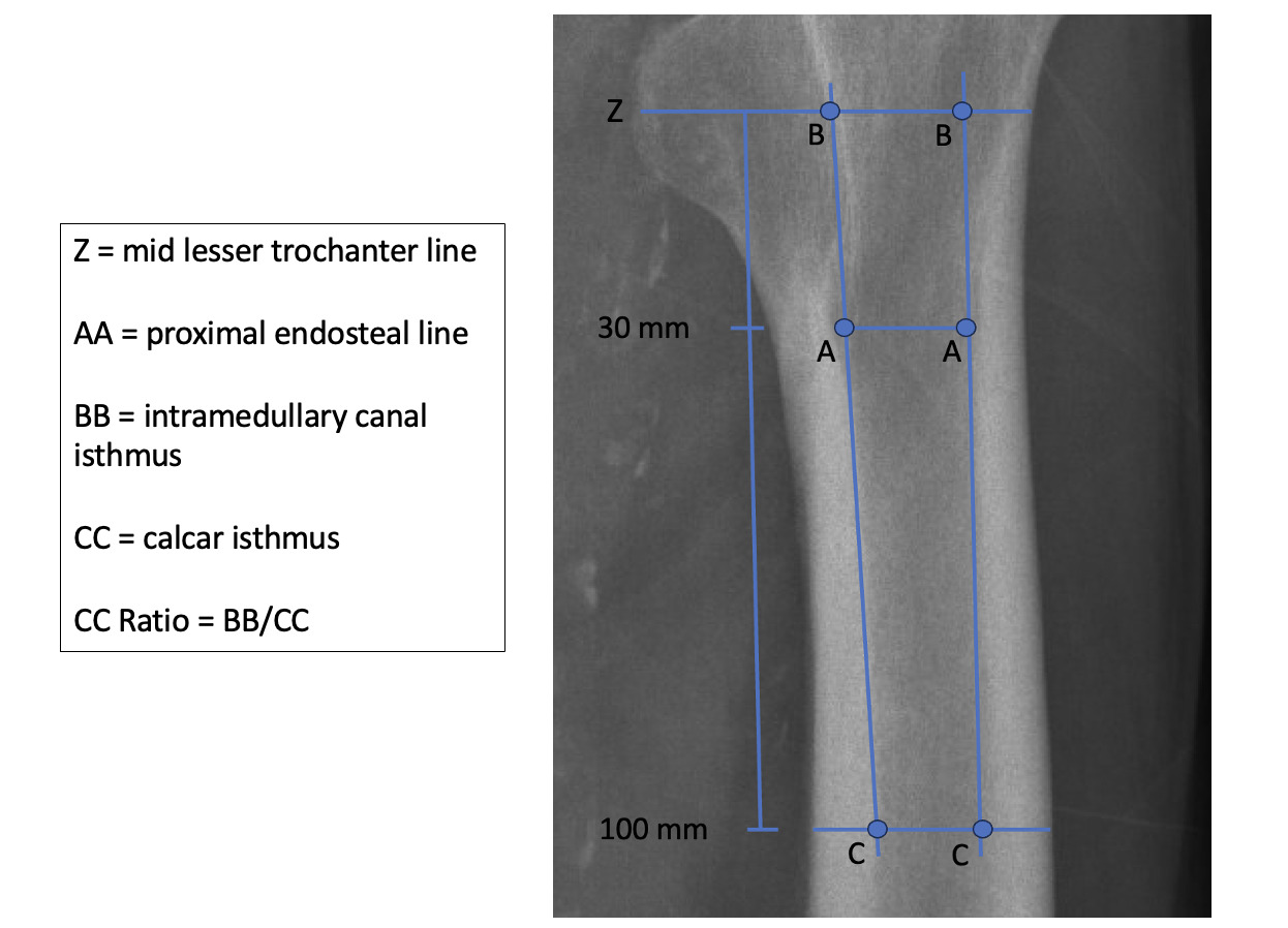
The primary endpoint of the study was the incidence of periprosthetic fracture. Patients were stratified based on proximal femur morphology using the Dorr classification system. Dorr classification was determined by measuring proximal femur dimensions using anterior-posterior (AP) hip radiographs taken at the time of femoral neck or head fracture [Figure 1]. A line was drawn through the midpoint of the lesser trochanter, perpendicular to the long axis of the femur. Longitudinal lines were then drawn through the canal width at 30 mm and 100 mm below the midpoint of the lesser trochanter. Intramedullary canal width was measured at 100 mm, and the calcar isthmus was measured using the longitudinal lines intersection point with the midpoint of the lesser trochanter. The intramedullary canal width was divided by the calcar isthmus to give the canal to calcar isthmus ratio. Canal to calcar isthmus (CC) ratios less than 0.5 were classified as having Dorr A proximal femur morphology, between 0.5 and 0.75 as having Dorr B morphology, and greater than 0.75 as having Dorr C morphology.
CTI was also calculated as a ratio of cortical thickness to total width at 100 mm below the midpoint of the lesser trochanter. Each of these assessments were performed by the primary author (LE), a medical student. Implant type, laterality of fracture, mechanism of injury, and American Society of Anesthesiologists (ASA) grade was recorded. The incidence of periprosthetic fracture was noted via chart review, and mechanism of periprosthetic fracture, time to fracture, intervention post fracture, and time to death were charted. Baseline and demographic characteristics were summed using descriptive statistics, and a t-test and chi-squared test was used to analyze the data. A p-value < 0.05 indicates statistical significance.
RESULTS
A total of 97 patients who underwent uncemented hemiarthroplasty within the study period were identified. The mean age was 79.1 years, and 60.9% were female (59/97). White patients represented 43.3% of the study population whereas 38.1% of patients were Black or African American, 12.4% were Hispanic or Latino, and 2.1% were Asian; the remaining four patients identified as unknown or other race. Across all patients that underwent an uncemented hemiarthroplasty, the most common comorbidities were dementia (24.7%) and diabetes (20.6%). 7.2% of patients were ASA grade 2, 79.4% were ASA grade 3, 12.4% grade 4, and 1.0% grade 5.
Nine patients (9.3%) were classified as having Dorr A proximal femur morphology, 62 patients (63.9%) as having Dorr B morphology, and 26 patients (26.8%) as having Dorr C morphology (Table 1). No Dorr A patients experienced a periprosthetic fracture. In total, three patients with Dorr B morphology at the time of injury and two patients with Dorr C morphology experienced a periprosthetic fracture (5.2%) [Table 1]. This result was not statistically significant (P=0.66). The most common comorbidities amongst this subset were osteoporosis (40%) and the presence of a solid tumor (40%). Osteoporosis, peripheral vascular disease, peptic ulcers, liver disease, chronic kidney disease, and the presence of a solid tumor were all more prevalent in patients that experienced periprosthetic fracture, although none achieved statistical significance (all P>0.05). Two of the fractures occurred intraoperatively during the hemiarthroplasty (on postoperative day 0), two during mechanical falls post-hemiarthroplasty (on postoperative days 64 and 2,172), and one during removal of hip prosthesis (on day 1,008 post-operatively). The mortality rate associated with the incidence of periprosthetic fracture was 40% (2/5), with a time to death of 4 days and 83 days following the second fracture event. The canal to calcar isthmus ratio was a mean of 0.68 across the entire patient population compared with 0.71 for the patients who experienced periprosthetic fracture (P=0.42). The mean CTI across all patients was 0.52, whereas the mean CTI across the patients that experienced periprosthetic fracture was 0.42 (P=0.006).
Of the 97 individual charts reviewed, the type of implant used in the uncemented hemiarthroplasty was identified in 95 of subjects. We found that 27 patients (28%) received a Zimmer Biomet (Warsaw, IN) implant, 35 (37%) a Stryker implant (Portage, MI), and 33 (35%) a DePuy Synthes (Raynham, MA) implant. Of the subset of patients who experienced a periprosthetic fracture, one (20%) patient had a Zimmer Biomet implant, two (40%) patients had a Stryker implant, and two (40%) patients had a DePuy Synthes implant. In comparison to the broader cohort, 3.7%, 5.7%, and 6.1% of patients who received these respective implants experienced a periprosthetic fracture.
DISCUSSION
In comparison to prior literature, a relatively low proportion of patients (9.3%) had Dorr A proximal femur morphology.17,18 Park et al. classified 56% of patients as Dorr A, however, the average age of the patients that demonstrated Dorr A morphology in their study was substantially younger than that of the present study (50.6 versus 79.1 years).17 In contrast, Murphy et al. describe similar distributions of proximal femur morphology, as 16% were Dorr A, 76.6% Dorr B, and 7.3% Dorr C.18 This closely aligns with our results and is likely attributable to patient populations more similar in age, as the cohort studied by Murphy et al. had a mean age of 80.4 years.18
Both Dorr B and Dorr C classifications were predominant in female patients. There were 37 female patients classified as Dorr B (60.0%) and 19 female patients classified as Dorr C (73.1%). In contrast, a higher proportion of male patient were denoted as Dorr A (66.7%). This distribution seems to align with previous literature as Wilkerson et al. found that older female patients have wider diaphyseal canals than younger female patients, a phenomenon which was not observed in male patients.12
With regard to proximal femur morphology, the highest incidence of periprosthetic fracture was seen in patients that have Dorr C classification (7.7%). This incidence is similar to the incidence of periprosthetic fractures found in the setting of uncemented hemiarthroplasty by Murphy et al. of 10.3%.18 As stated previously, patients with Dorr C proximal femur morphology have wider femoral canals and thinner cortices as compared to those that have Dorr B and Dorr A proximal femur morphology.12 It is important to note, however, that no significant differences in periprosthetic fracture risk incidence were observed across the three Dorr classifications.
Furthermore, the cortical thickness index, which is another radiographic measurement that can help assess bone morphology, was on average 0.52 for all patients that were reviewed, but just 0.42 for patients that experienced periprosthetic fracture. This corresponds with an approximately 19% lower index, which was statistically significant. Therefore, this result indicates that patients who developed periprosthetic fracture had thinner, potentially compromised, cortices and supports using CTI to assist in evaluating risk of periprosthetic fracture prior to surgery. This result contrasts with the existing literature, as CTI demonstrated no association with femoral stem subsidence according to Badoux et al.15 Further, a retrospective analysis of 233 patients who underwent endomedullary nailing or total/hemiarthroplasty for the treatment of proximal femur fracture found no correlation with CTI and mechanical complications, including periprosthetic fracture.16 Regardless, CTI should not be considered in isolation from other factors, as patients with femoral head or neck fractures present with varying comorbidities, ages, and levels of activity, which all play a role in overall outcomes. Nonetheless, our research highlights the potential importance of evaluating proximal femur morphology to assess the risk of periprosthetic fracture following uncemented hemiarthroplasty.
The study was limited by its overall sample size (97 patients), as well as the number of patients who experienced a periprosthetic fracture (5 patients). This is likely to contribute to study underpowering and increases the probability of Type II error as no statistically significant difference in periprosthetic fracture risk across the three Dorr classifications was observed. Additionally, this study was retrospective in nature, therefore there is inherent bias. Assignment of Dorr classification and calculation of CTI by a single individual precluded interobserver reliability; intraobserver reliability testing was also not performed. Lastly, patients may have experienced fractures outside of our institution and would not have been captured as an outcome.
CONCLUSION
This retrospective study found a nonsignificant higher incidence of periprosthetic fracture following uncemented hemiarthroplasty in patients who had Dorr C proximal femur morphology than those with Dorr A and Dorr B classifications. In contrast, the mean cortical index amongst patients who developed periprosthetic fracture was significantly lower than the comparison group. Put together, these findings demonstrate that cortical index may confer greater utility than Dorr classification when evaluating the risk of periprosthetic fracture amongst patients eligible for uncemented hemiarthroplasty.